New gene-editing technique holds potential for treating childhood blindness

September 29, 2023
Share

Using a new experimental technique to fix faulty eye cells, a team led by UW–Madison researchers was able to repair a gene mutation that causes one form of childhood blindness. The scientists showed that their approach worked in lab-grown cells derived from a patient with the currently untreatable inherited disease Leber congenital amaurosis (LCA) and a mouse model that mimics the disease.

The method hinges on delivering gene-editing tool CRISPR base editor to retinal cells using silica nanocapsules. The proof-of-concept study restored the function of a protein that controls the flow of potassium ions in retinal tissue, which allows light-detecting cells to work properly.

“Our goal is to design a package that will carry CRISPR base editors to the retina,” said Bikash Pattnaik, co-investigator and associate professor of pediatrics at the UW School of Medicine and Public Health. “It will be able to introduce nanoparticles in the eye, and those nanoparticles will be designed to target the cell types identified for therapy.”

Left to right: Bikash Pattnaik, Meha Kabra and Pawan Shahi

Pattnaik, who holds a doctorate in biophysics and is affiliated with the Department of Ophthalmology and Visual Sciences, led a team including scientists and engineers from UW–Madison, Harvard, and MIT that published the study in the Journal of Clinical Investigation. The researchers are seeking to develop treatments for two forms of childhood blindness caused by retinal gene mutations, LCA and Best disease, which is also known as Best vitelliform macular dystrophy. LCA begins in infancy and causes extreme farsightedness, sensitivity to light, and involuntary eye movements. Best disease, usually diagnosed in childhood, causes macular degeneration and loss of central vision, visual acuity, and color perception.

The diseases are relatively rare, affecting 3 to 4 individuals per 100,000 people, but the research team expects that its new nanoparticle-packaged CRISPR technology will be able to treat other inherited eye diseases.

An innovative technique distinguishes the work from a more typical method used in gene editing. Since the CRISPR gene editing method known as CRISPR/Cas 9 was discovered in 2012, using a modified virus has become the norm for delivering gene-correcting material. However, the virus transport method has disadvantages in its degree of precision, the number of cells affected, and the possibility of unpredictable side effects.

The team reasoned that silica nanocapsules do not interact with the body, lowering the risk of problematic and unpredictable immune system responses that viral methods may elicit. Another advantage of the technique is its specificity. “The rapid pace of genetic diagnosis development has enabled accuracy in identifying disease-causing mutations, and we used base editing to correct a specific error in the DNA sequence of patients, overcoming several challenges through the collaborative team science approach,” said research associate Meha Kabra.

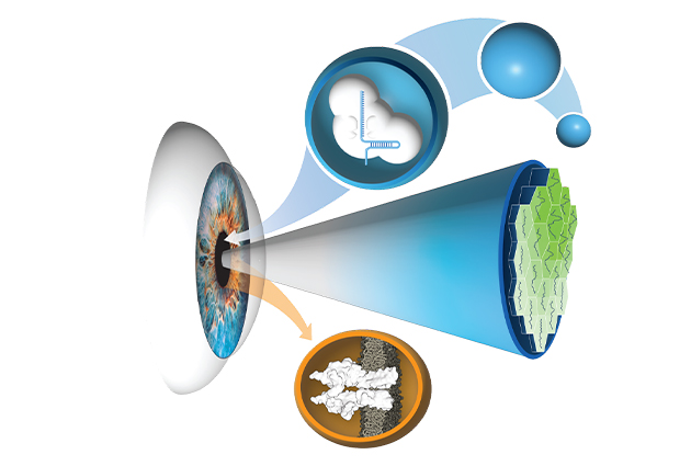
A diagram showing how an eye works
In an experimental technique, researchers delivered genome editors to the eye to correct a gene deficit that causes Leber congenital amaurosis, rescuing vision in an animal model.
Image by Bikash Pattnaik

Showing effectiveness in a living organism was complicated because mice with two copies of the LCA-causing mutation do not survive past infancy, whereas two copies of homozygous mutations in humans are not lethal. The researchers generated mice with one copy of the mutation, then used gene editing to disrupt the second normal copy in retinal cells to enable testing of the therapy. “The clinical relevance and efficacy of any potential new therapy requires studies using laboratory animal models,” said scientist Pawan Shahi. “Without an existing animal model for this specific form of blindness, we used genome editing to first create the model and then deliver the base editor and demonstrate therapeutic value.”

“Typically, drug development can take more than 30 years,” Pattnaik said. “But with a multidisciplinary approach that brings together people with different expertise, we can cut this timeline significantly.” He says that this work, which demonstrates the feasibility of repairing genetic defects in ion channel proteins, is a critical first step toward restoring the sight of affected young patients.

Funding for the study was provided by the UW–Madison Stem Cell and Regenerative Medicine Center for M. Kabra; support from the Retina Research Foundation for K. Saha, B. Pattnaik, and D. Gamm; support from the McPherson Eye Research Institute for D. Gamm; the Wisconsin Partnership Program; the Harrington Discovery Institute (K. Saha); the Helen Hay Whitney Foundation; Howard Hughes Medical Institute; National Institutes of Health K99 HL163805, NIH R01 EY024995, NIH R24 EY032434, NIH U01 AI142756, NIH RM1 HG009490, and NIH R35 677 GM118062; and funds from Research to Prevent Blindness. The study was partly supported by S10OD018221 and NIH P30 EY016665.